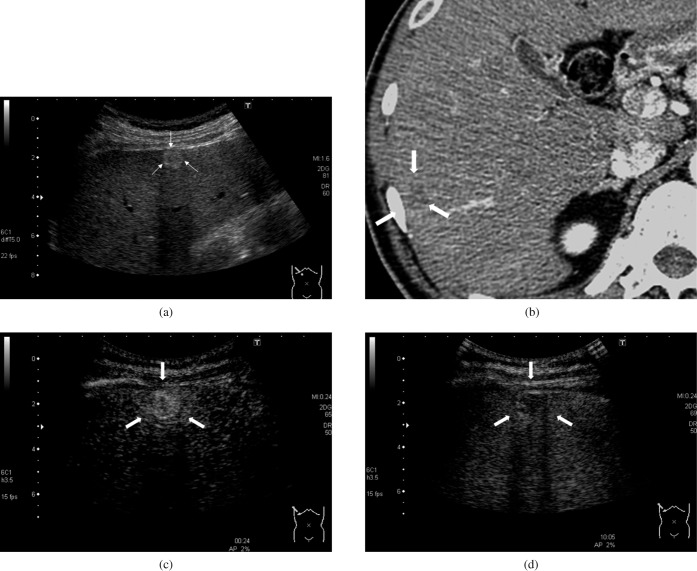
Figure 1.
A 52-year-old man with hepatitis C-related cirrhosis and moderately differentiated hepatocellular carcinoma (15.2 mm). (a) B-mode sonogram. A hyperechoic lesion was observed in the liver (arrows). (b) Contrast-enhanced CT, hepatic artery-dominant phase. The lesion showed a hypovascular appearance on the CT image (arrows). (c) Contrast-enhanced ultrasound, early phase. The intensity difference between the hepatic lesion and adjacent liver parenchyma was 22.6 dB, which was higher than the maximum intensity difference in the non-tumour parenchyma (2.0 dB). The lesion appeared as hyperenhanced (arrows). (d) Contrast-enhanced ultrasound, late phase. The intensity difference between the hepatic lesion and adjacent liver parenchyma was –6.6 dB, with the absolute value greater than the maximum intensity difference in the non-tumour parenchyma (2.1 dB). The lesion appeared as hypoenhanced lesion (arrows).

