
Fig. 2.
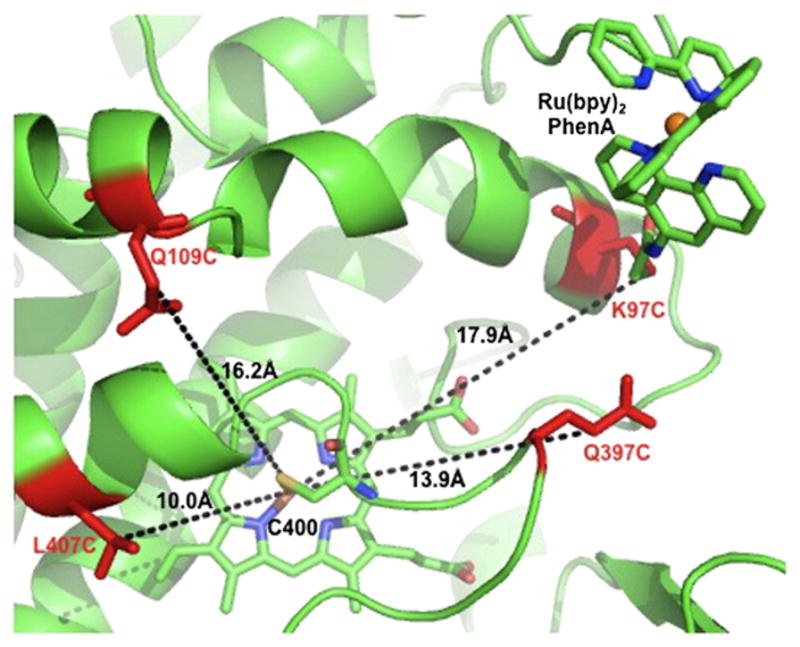
Cartoon representation of the mutated residues Q109C, K97C, Q397C and L407C (red) in the series of hybrid enzymes adapted from the K97C-1 crystal structure (PDB ID: 3NPL). The estimated distances from the Cβ carbon of the side chain residue to the Fe center are indicated.