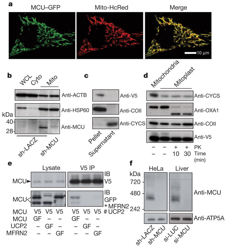
Figure 3. MCU is oligomeric and resides in the mitochondrial inner membrane as a larger complex.
a, Confocal imaging of MCU–GFP co-expressed with mitochondria-targeted HcRed (Mito-HcRed) in HeLa cells. b, Immunoblot analysis of HeLa whole-cell lysate (WCL), cytosol (Cyto) or crude mitochondrial fractions (Mito), using antibodies against MCU, HSP60 (matrix protein, also known as HSPD1), or ACTB (cytosol). c, Immunoblot analysis of soluble (supernatant) and insoluble (pellet) fractions following alkaline carbonate extraction of mitochondrial fractions from HEK-293 cells expressing MCU–V5. Immunoblot analysis was performed using antibodies against V5, COII (integral inner membrane protein) and CYCS (soluble intermembrane space protein). d, Immunoblot analysis after proteinase K (PK) treatment of MCU–V5-expressing HEK-293 mitoplasts for indicated times. e, Anti-V5 immunoprecipitations performed as in Fig. 1d using lysates from HEK-293 cells stably expressing MCU–V5 and transiently transfected with MCU–GFP, UCP2–GFP, or MFRN2–GFP. f, Blue native PAGE analysis of mitochondrial fractions from HeLa cells (stably expressing sh-LACZ or sh-MCU, left panel) or from livers of mice (si-LUC or si-MCU, right panel) and immunoblotted for MCU. ATP5A1 is used as a loading control.

