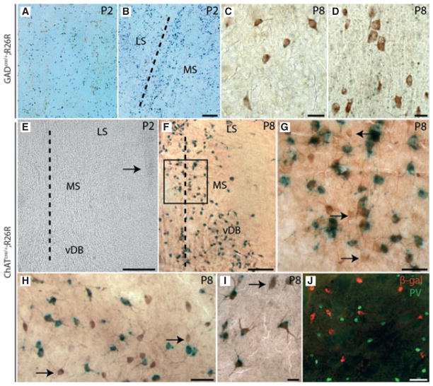
Fig. 1.
Cre-recombination in the subcortical telencephalon of GADcre/+;R26R and ChATcre/+;R26R mice. (A–D) X-gal staining is shown for GADcre/+;R26R mice at P2 (A, B) and P8 (C, D). As shown here by representative coronal sections, an intense punctuate-like labelling was observed in newborn animals throughout the telencephalon including the CPu (A) and within the septal complex (B). The border between the medial septum (MS) and the lateral septal complex (LS) is marked by a dotted line (B). From P7–8 onwards, these precipitates could be localized in GABAergic and cholinergic neurons. As shown here for the CPu (C) and the MS (D), nearly all of the ChAT-ir neurons of the ventral forebrain were also found to be positive for X-gal. Scale bars – 50 μm (A, B), 30 μm (C), 20 μm (D). (E–J) In ChATcre/+;R26R mice, no labelling for X-gal was observed prior to or shortly after birth in the telencephalon. A corresponding coronal section obtained at P2 is shown (E). The ventricle located lateral to the various septal nuclei (MS, vDB, LS) is marked by an arrow. The dotted lines (E, F) indicate the median axis of the brain. (F–J) At P8, intense green punctuate-like labelling for X-gal was observed in all cholinergic centres of the ventral forebrain including the MSvDB (F; boxed area is shown at higher magnification in G), the magnocellular complex of the hDB-SI (H), and in the CPu (I). In these regions, the overwhelming majority of X-gal-positive cells could be identified as cholinergic neurons following subsequent IHC for ChAT (brown labelling of cells; F–I). Only a small subset of ChAT-ir neurons did not present any labelling for X-gal (marked by arrows in G–I). In ChATcre/+;R26R mice, activity of Cre recombinase was found to be restricted to cholinergic neurons. As shown for the CPu, no co-localization of the two markers β-gal (red cells) and PV (green cells) could be detected in the ventral forebrain (J). Scale bars –100 μm (E, J), 200 μm (F), 50 μm (G, H) and 10 μm (I).

