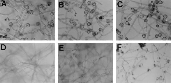
Figure 1.
Light-microscopic photographs of pollen tubes grown in vitro with different carbon sources in 2% Suc after 8 h (A) and 24 h (D); in 2% Fru after 8 h (B) and 24 h (E) (pollen tubes grown in 2% Glc revealed an identical picture (not shown); and in 2% mannitol after 8 h (C) and 24 h (F). Pollen tubes in D and E are out of focus because they float in the medium due to their length. A through C, Magnifications ×100; D through F, magnifications ×64.

