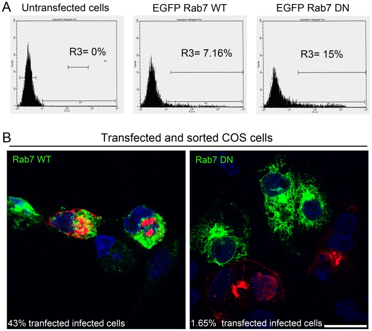
Figure 4. Late endosomal compartment relevance for ASFV infection.
(A) Representative FACS profiles obtained during sorter analysis of COS-7 cells transfected with GFP-Rab7-wild type (Rab7 WT) and dominant negative mutant (GFP-Rab7-DN, T22N). R3 represents transfected cells expressing GFP to be sorted. (B) Representative confocal micrographs of transfected, sorted cells after isolation, infected with ASFV at a moi of 1 for 24 hpi and immunostained for major viral capsid protein p72 (red). Percentages of transfected infected cells decreased from 43.5% in cells expressing Rab7 WT to 1.65% in cells expressing Rab7 DN. Bar 25 µm.

