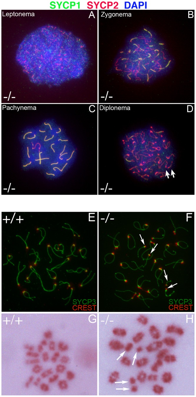
Figure 7. Formation of univalent chromosomes in Chtf18 −/− spermatocytes.
Meiotic chromosome spreads from Chtf18 −/− juvenile males at postnatal day 21 were stained by immunofluorescence with anti-SYCP1 serum (green), anti-SYCP2 serum (red), and DAPI (blue) (A–D). SYCP1 localizes to central elements of the synaptonemal complex and SYCP2 localizes to the axial/lateral elements of the synaptonemal complex. The leptone, zygotene, pachytene, and diplotene stages of prophase I Chtf18 −/− spermatocyte (−/−) are shown (A, B, C, and D respectively). Arrows in D reveal the presence of univalent chromosomes in the diplotene stage of prophase I. Wild-type and Chtf18 −/− spermatocytes from juvenile males at postnatal day 21 (E and F, respectively) were stained with CREST autoimmune serum, which stains centromeres, and anti-SYCP3, which stains the axial/lateral elements of the synaptonemal complex. Arrows in F indicate univalent chromosomes in a Chtf18 −/− diplotene spermatocyte. Metaphase I spread analysis of spermatocyte nuclei from juvenile males at postnatal day 21 stained with Giemsa (G and H). Twenty bivalent chromosomes are present in the wild-type metaphase I spermatocyte (G). A Chtf18 −/− metaphase I spermatocyte (H) demonstrates the presence of four univalent chromosomes (arrows in H).

