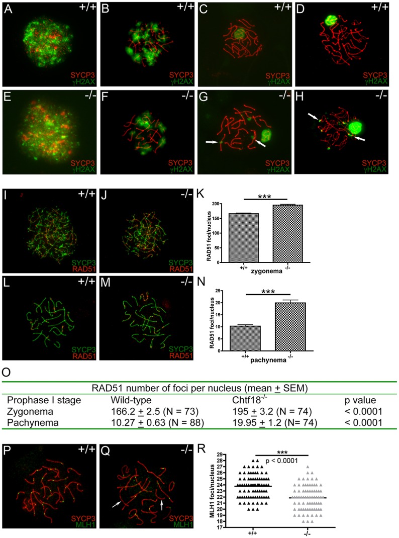
Figure 8. Meiotic recombination is defective in Chtf18 −/− spermatocytes.
Meiotic chromosome spreads from wild-type (A–D) and Chtf18 −/− (E–H) juvenile males at postnatal day 14 or 21 were stained with anti-SYCP3 (red) and γH2AX (green). A & E represent leptonema; B & F represent zygonema; C & G represent pachynema; D & H represent diplonema. Arrows in G and H show persistence of γH2AX in Chtf18 −/− spermatocytes. Meiotic chromosome spreads from wild-type (I and L) and Chtf18 −/− (J and M) males were stained with anti-SYCP3 (green) and anti-RAD51 (red). I and J represent zygonema; L and M represent pachynema. RAD51 focus counts per nucleus in zygonema (K and O) and pachynema (N and O) are shown. Meiotic chromosome spreads from wild-type and Chtf18 −/− spermatocytes were stained with anti-SYCP3 (red) and anti-MLH1 (green) (P and Q). Arrows indicate homologues without MLH1 foci. MLH1 focus counts per nucleus are shown in R.

