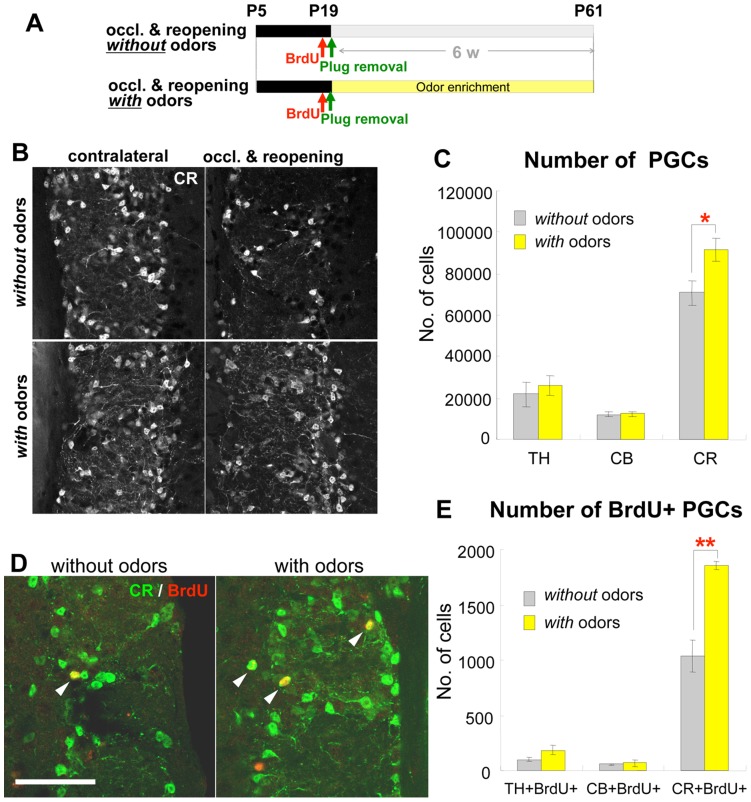
Figure 3. Odor stimulation recovers the reduction in CR+ PGCs caused by transient neonatal olfactory deprivation. A.
: Experimental procedure. Mice undergoing 2-week hemilateral naris occlusion from P5 were injected with BrdU just before naris reopening at P19 by removal of the nasal plug, then exposed to 21 different odorants that were changed every day for 6 consecutive weeks (with odors) or left in the normal breeding conditions without odor presentation (without odors). Histological analyses were performed at P61. B–C: Effect of odor stimulation on PGC populations. Coronal sections of the glomerular layer of P61 mice of each group, immunostained for CR (B). The number of CR+ PGCs, but not of TH+ or CB+ PGCs was significantly increased in mice exposed to odors (with odors, B, bottom panels; C, yellow bars, n = 5) compared with mice not exposed to presented odors (without odors, B, top panels; C, gray bars, n = 6). *P<0.05 D–E: Effect of odor stimulation on the addition of PGCs. Coronal sections of the glomerular layer of P61 mice of each group, immunostained for BrdU (red) and CR (green) (E). Arrowheads in E indicate BrdU+CR+ cells. The number of BrdU+CR+ PGCs, but not of BrdU+TH+ or BrdU+CB+ PGCs, was significantly increased in mice exposed to odors (with odors, D, right panel; E, yellow bars, TH and CB: n = 3, CR: n = 4) compared with those in mice without odor presentation (without odors, D, left panel; E, gray bars, TH and CB: n = 3, CR: n = 4). **P<0.01 Scale bars: 50 µm.

