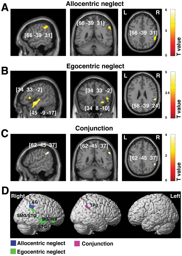
Figure 3. Voxel-wise statistical analysis of grey matter damage: allocentric vs. egocentric neglect at the chronic phase following stroke.
VBM results showing voxels corresponding to grey matter damage in (A) left allocentric, (B) left egocentric and (C) both forms of neglect (conjunction analysis). Please note that in A, B and C the lesioned areas are coloured according to their significance level in the VBM analysis, where a brighter colour indicates a higher t-value. The numbers in brackets indicate the peak MNI coordinates. (D) To further illustrate the relationship between grey matter loss and any associated allocentric or egocentric symptoms at the chronic phase, all clusters identified by VBM as described above are plotted on a rendered brain. AG, angular gyrus; BG, basal ganglia; INS, insula; SMG, supramarginal gyrus; TPJ, temporal-parietal junction.

