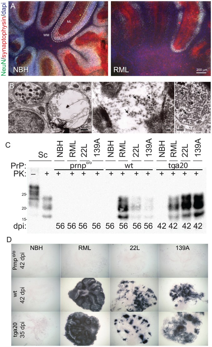
Figure 1. Localization and consequences of prion replication in COCS.
(A) Fluorescence micrographs showing profound loss of NeuN+ cerebellar granule cells and of synaptophysin in wt COCS challenged with RML prions (56 dpi, right panel). No neuronal damage was detected in COCS exposed to non-infectious brain homogenate (NBH; left panel). WM: white matter. ML: molecular layer. CGL: cerebellar granule cell layer. (B) Electron microscopy showing membrane-enclosed intraneuronal spongiform vacuoles (left), tubulovesicular structures (PrP-deficient spheres measuring between 20 and 40 nm in diameter, arrow, middle), and degenerating axons accumulating intra-cellular organelles including mitochondria (arrow, right) in RML-infected wt slices at 56 dpi. (C) Immunoblots showing PrPSc in tga20 and wt, but not Prnpo/o COCS exposed to prions (RML, 22L, 139A) or NBH. Sc; scrapie-sick wt mouse brain homogenate, used as positive control. (D) Histoblots showing strain-specific differences in PrPSc deposition patterns of tga20 and wt COCS. No PrPSc signal was observed in Prnpo/o COCS and in PrP-expressing COCS exposed to NBH.

