
Figure 1. Electrophysiological recordings confirm the location for in vivo injection of retrograde tracers.
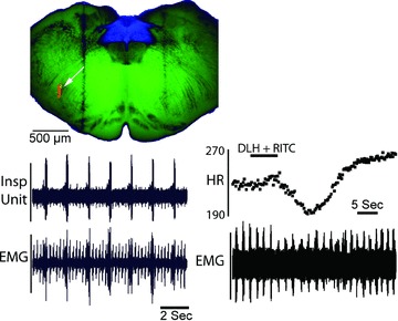
Respiratory responses to a combined injection of rhodamine latex beads (RITC) and dl-homocysteate (DLH) into the rostral VRG. Upper left panel, transverse slice used for in vitro recording of NTS neurons and showing the injection site in the rVRG (arrow). Lower left panel, rVRG inspiratory unit activity (upper trace) recorded in vivo immediately prior to the RITC/DLH injection. Lower trace is respiratory muscle EMG and ECG recording. ECG potentials were graphically attenuated to enhance the respiratory EMG. Lower right panel, heart rate (upper trace, instantaneous frequency in beats min−1) and respiratory EMG (lower trace) responses to a small injection (3 nl) of DLH and rhodamine latex beads as part of a total injection volume of 42 nl. Subsequent injection of a larger portion of the incremental injection produced a sustained apnoea and more marked bradycardia. The 300 μm thick medullary slice was captured as a brightfield image and light–dark inverted in Photoshop. Brightness and contrast for one copy of this image was adjusted to optimally show the myelinated fascicles in the green channel of a blank RGB image. A second copy was adjusted to optimize the NTS and was pasted in the blue channel of the RGB image. The epifluorescence image of the RITC injection was inserted into the red channel of the same RGB image. Dark vertical bands are the result of the nylon retaining bands in the in vitro chamber.