
Figure 4. Astrocyte-driven action potentials in inspiratory neurons.
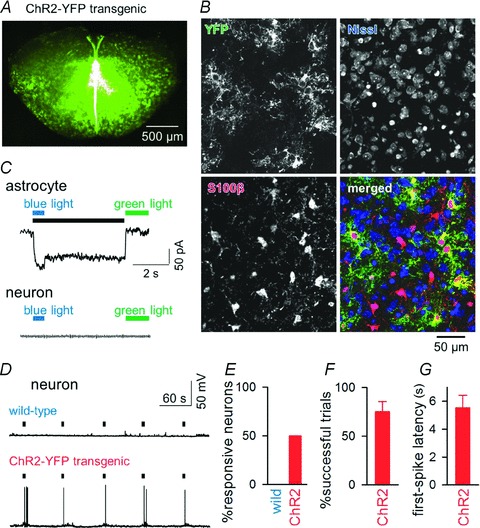
A, YFP fluorescence of a rhythmically active slice prepared from a Mlc1-tTA::tetO-ChR2(C128S)–YFP transgenic mouse. B, triple labelling of preBötC cells with an antibody against green fluorescent protein (i.e. YFP appearing in green) to indicate ChR2-expressing cells, with fluorescent Nissl to show neuronal somata (blue), and with an antibody against S100β to identify astrocytes (red). ChR2–YFP positivity was found exclusively in S100β-positive cells. Thus, ChR2 was expressed only in astrocytes. C, switch-on (blue light) and switch-off (green light) of a persistent inward current in a voltage-clamped ChR2-expressing astrocyte (top) and neuron (bottom). Voltages were clamped at −70 mV. The black bar indicates the activated period. D, photo-activation of astrocytes triggers action potentials in an inspiratory neuron from the preBötC in a ChR2(C128S)YFP transgenic (but not in a wild-type) mouse. Black bars indicate the activation periods of preBötC astrocytes. E, ratios of neurons that fired in response to photo-activation of astrocytes in wild-type (n= 9 neurons) and in ChR2(C128S)YFP transgenic mice (n= 16 neurons from 5 animals). F, ratios of the trials in which light stimulation elicited action potentials (n= 8 responsive neurons). G, latency from the light-stimulus onset to the first spike (n= 8 neurons).