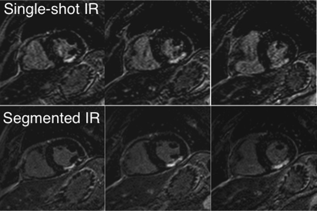
Figure 4.
Short-axis view images of a 64-year-old patient after myocardial infarction with a predominantly transmural scar of the inferior wall. The images in the upper row were acquired with the single-shot IR–GE technique; the images in the lower row with the segmented 2D IR–GE technique served as a reference technique. 2D, two-dimensional; GE, gradient echo; IR, inversion recovery.

