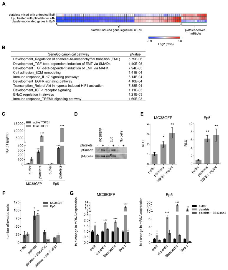
Figure 2. Platelet-Induced Gene Expression Signature Reveals Increased Expression of Prometastatic Genes and Activation of the TGFβ Pathway in Tumor Cells.
(A) Heat map of genes regulated by more than 4 fold (p<0.05) in Ep5 cells treated with platelets in comparison with untreated Ep5 cells (line 2). Line 1 and line 2 show Log2 ratios of gene expression compared to untreated Ep5 cells. mRNAs present in platelets (line1) were removed from the list of genes modulated upon platelet treatment of Ep5 cells (line 2) to generate a platelet-induced gene signature (line 3), which is listed in Table S2.
(B) Canonical signaling pathways most significantly associated with the list of genes differentially expressed by Ep5 cells upon platelet treatment (platelet-induced gene signature; Table S1; threshold = 2 fold, up and down regulated genes considered, p<0.05) as determined with GeneGo canonical pathway maps. The ten pathways with the lowest p values are shown.
(C) Concentration of active and total TGFβ1 in conditioned medium from MC38GFP or Ep5 cells treated with buffer or platelets for 40h. The conditioned medium was collected, centrifuged to remove platelets, and the presence of TGFβ1 in the supernatant measured by ELISA. Each bar represents the mean ± SEM of n=2–6. ***p<0.001 as determined by Student’s t-test.
(D) Detection of phospho-Smad2 protein levels by immunoblotting of Ep5 or MC38GFP cells treated as in (C). Amounts of platelets equal to those used to treat cells were also loaded as control (no cells). β-tubulin is used as loading control.
(E) Relative luciferase activity (RLU) in MC38GFP or Ep5 cells stably expressing a luciferase reporter under the control of an SBE promoter and treated for 40h with buffer, platelets or 1ng/ml TGFβ1 (positive control) (n=5–6).
(F) MC38GFP and Ep5 cells were added at the top of transwells coated with Matrigel and treated with buffer, platelets, platelets + SB431542 (10μM) or platelets + TGFβ1 blocking antibody (6μg/ml). The total numbers of cells that invaded to the bottom of the transwell were counted after 48h (n=3).
(G) Relative fold change in mRNA expression in MC38GFP or Ep5 cells treated with buffer, or platelets +/− SB431542 (10μM) for 40h (n=3). Values are normalized to Gapdh expression.
For panels E, F and G bars represent the mean ± SEM, and *p<0.05, **p<0.01, ***p<0.001 vs buffer were determined by one-way ANOVA followed by Tuckey’s post test.

