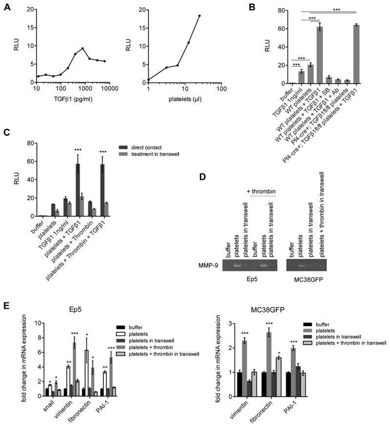
Figure 5. TGFβ1 and Direct Platelet-Tumor Cell Contact Synergize to Promote Prometastatic Gene Expression.
(A) Relative luciferase activity (RLU) in MLEC cells stably expressing a luciferase reporter under the control of a TGFβ responsive PAI-1 promoter construct and treated with different concentrations of TGFβ1 (left panel) or platelets (right panel). Note that the y-axis scale is the same for both panels and that platelets give higher stimulation than achievable with TGFβ1 alone.
(B) Relative luciferase activity (RLU) in MLEC cells stably expressing a luciferase reporter under the control of a PAI-1 promoter construct treated with buffer, platelets from WT or Pf4-cre+; TGFβ1fl/fl mice, TGFβ1 1ng/ml or with combinations of platelets + TGFβ1 1ng/ml, +/− SB431542 (SB; 10μM) or +/− TGFβ1 blocking antibody (Ab; 6μg/ml)(n=3–16).
(C) Relative luciferase activity (RLU) in MLEC cells stably expressing a luciferase reporter under the control of a PAI-1 promoter construct treated with buffer, platelets, or thrombin-activated platelets +/− 1ng/ml TGFβ1 seeded either at the bottom (direct contact with tumor cells) or in the upper chamber of a transwell (0.4μm pore size) to prevent direct contact between platelets and tumor cells (n=2–3).
(D) Zymography for MMP-9 in the conditioned medium from Ep5 or MC38GFP cells treated with buffer, platelets, or thrombin-activated platelets seeded either at the bottom or in the upper chamber of a transwell (0.4μm pore size).
(E) Relative fold change in mRNA expression in Ep5 or MC38GFP cells treated as in (D) (n=3). Values are normalized to Gapdh expression.
For panels B, C and E, bars represent the mean ± SEM, and *p<0.05, **p<0.01, ***p<0.001 vs buffer (unless otherwise indicated) were determined by one-way ANOVA followed by Tuckey’s post test.

