
Figure 3.
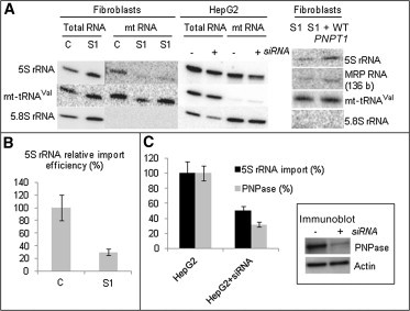
Quantification of Mitochondrial Import of 5S rRNA and MRP RNA by RNA Hybridization
(A) On the left, total and mtRNA from cultured skin fibroblasts of a control (C) and subject 1 (S1) were separated in denaturing 10% PAGE, transferred on Hybond-N filters, and hybridized with oligonucleotide-labeled probes for 5S rRNA, mt-tRNAVal, and 5.8S rRNA. Two deposits (1 and 3 μg) of subject mtRNA were analyzed for 5S-rRNA-import analysis. In the middle is Northern blot hybridization with 5S-rRNA-specific, mt-tRNAVal-specific, and 5.8S-rRNA-specific radio-labeled probes of total and mtRNA from HepG2 cells transfected or not with PNPT1 siRNAs. On the right, Northern blot analysis of mtRNA after transient expression of WT PNPT1 cDNA restored both 5S rRNA and 136 bp MRP RNA amounts in the mitochondria of subject 1 (S1) fibroblasts.
(B) Relative values of 5S rRNA import in subject 1 (S1) fibroblasts normalized to the control (C). Error bars correspond to two independent experiments.
(C) Relative 5S rRNA import and PNPase amount in control HepG2 and treated HepG2 + siRNA cells. The error bars correspond to two independent experiments. Immunoblot analysis of PNPase downregulation is shown in the inset. The actin-specific antibody was used as a loading control.