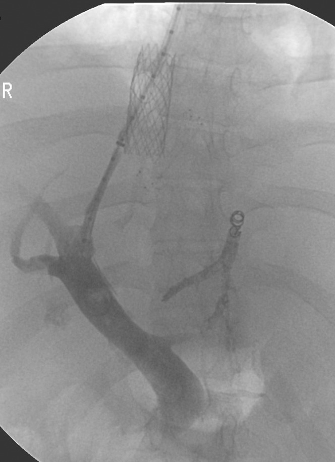
Figure 2.

A royal flush pigtail catheter tip seen in the portal vein through the inferior vena cava (IVC) strut and parenchymal track opacifies the portal vein. The dilated strut can be seen clearly in the IVC stent. The previously deployed accessory hepatic venous stenting can also be seen. A branching column of contrast with coils at the upper end represents a previous intervention attempted 1 week previously (percutaneous left hepatic vein access with failed access into the IVC owing to long segment stenosis and narrow ostium).
