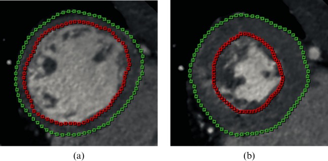
Figure 1.
A 63-year-old woman with atypical chest pain. Example of 64-slice MDCT short-axis reconstructions in (a) end-diastole and (b) end-systole to assess global left ventricular (LV) function. LV endocardial and epicardial contours drawn on reformatted short-axis views show that papillary muscles and trabeculae are included in the ventricular volume. The example is provided of a patient with a normal LV ejection fraction of 62%.

