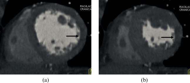
Figure 3.
A 73-year-old man with a history of lateral myocardial infarction with a heart rate of 70 beats per minute. The multidetector CT short-axis images at (a) end-diastole and (b) end-systole disclose akinesia in the lateral region (arrows). The example is provided of a patient with a normal left ventricular ejection fraction of 61%.

