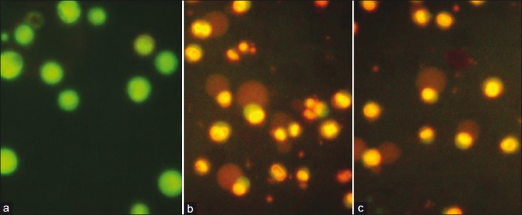
Figure 3.

DLA cells stained with acridine orange–ethidium bromide (Ao-Et Br) under a fluorescent microscope. (a) DMSO control DLA cells appeared in yellowish green color (live). (b) The active fraction (25 μg/mL) of Z. diphylla-treated DLA cells showing cell death (orange-red) and apoptotic bodies. (c) Curcumin (25 μg/mL)-treated DLA cells showing cell death (orange-red) and apoptotic bodies
