Figure 3.

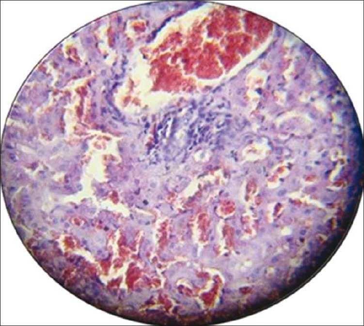
Photomicrograph of the liver section with moderate histological findings (portal tract dilatation and congestion inflammatory cell infiltration, sinusoidal dilatation congestion). Stained with hematoxylin and eosin (×40)

Photomicrograph of the liver section with moderate histological findings (portal tract dilatation and congestion inflammatory cell infiltration, sinusoidal dilatation congestion). Stained with hematoxylin and eosin (×40)