
Figure 3.
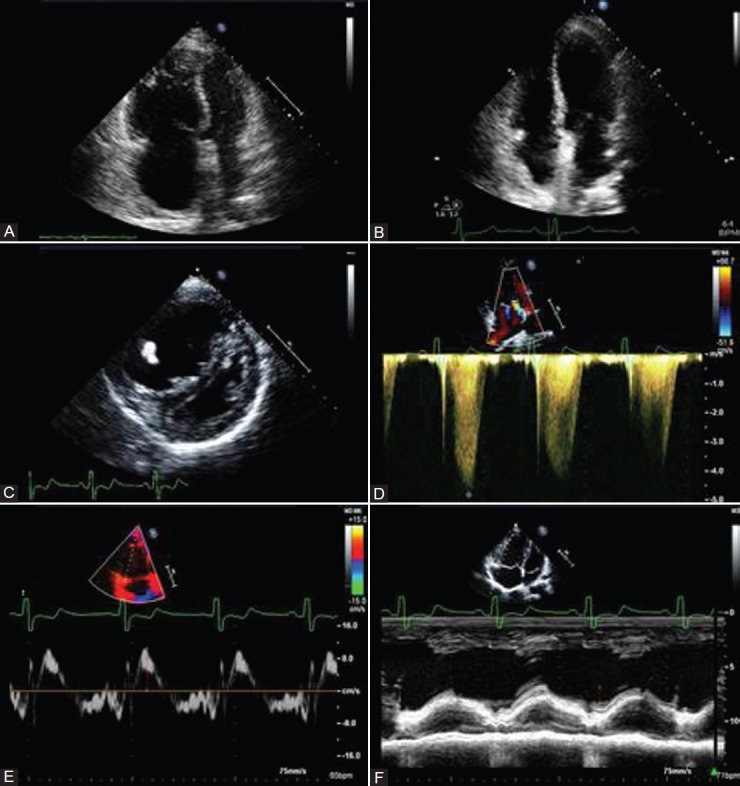
(A) RV enlargement and hypertrophy in patient with CTEPH. (B) Normal RV size 6 months after PTE in the same patient as in A. (C) Parasternal short-axis view demonstrating RV dilation and hypertrophy and displacement of septum. (D) Tricuspid regurgitation jet demonstrating severe PH in patient with PAH. (E) Peak systolic velocity of tricuspid annulus borderline depressed at 10 cm/s. (F) TAPSE in patient with PAH measured at 14 mm consistent with RV hypokinesis.