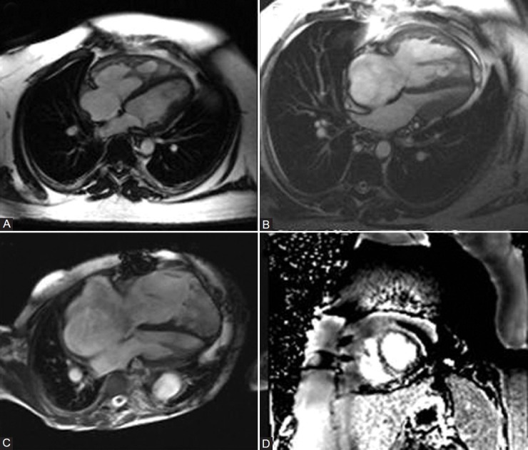
Figure 4.

(A) Mild RV and RA dilation with RV hypertrophy and prominent hypertrophy of moderator band. (B) Severe RV and RA dilation with RVH and compression of left-sided chambers by septal deviation. (C) End-stage RV failure with marked RV and RA dilation and thinned out RV free wall. (D) Late gadolinium-enhanced short-axis image demonstrating fibrosis at the RV septal insertion point.
