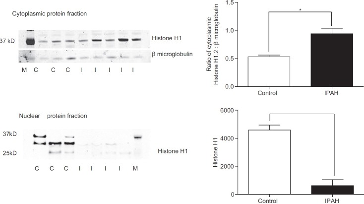
Figure 6.
Representative western blots of cytoplasmic and nuclear fractions of control and IPAH PASMCs. Histone H1 expression was significantly increased in the IPAH cells compared to controls. Nuclear histone H1 expression was significantly reduced in the IPAH nuclear fraction compared to controls. Densitometric data for histone H1 is related to ß microglobulin. A total of three control different cell lines and four IPAH cells lines were examined. Data are shown as mean + SEM; *P < 0.05.

