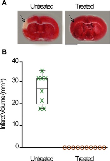
Figure 3.

Stimulation treatment protects cortical structure in aged rats. A, Representative coronal sections from an untreated (left) and a treated rat (right) with TTC assay for infarct. Red staining indicates healthy tissue; lack of staining indicates ischemic infarct. Arrows point toward MCA blood supply territory. Scale bar = 5 mm. B, Box-and-whisker plot of infarct volume (mm3), with individual subjects plotted for untreated (green) aged subjects and treated (gold) aged groups.
