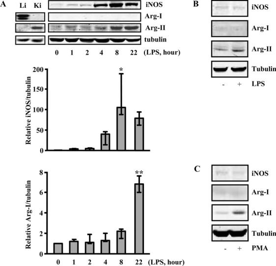
Figure 1.

Upregulation of Arg‐II but not of Arg‐I in response to inflammatory activation and differentiation of monocytes/macrophages. A, The murine macrophage cell line RAW264.7 was stimulated with LPS (0.1 μg/mL) for the indicated time points (hours). The expression of iNOS, Arg‐I, and Arg‐II was detected by Western blot. Protein lysates of murine liver (Li) and kidney (Ki) were used as positive controls for Arg‐I and Arg‐II, respectively. Representative blots from 3 independent experiments are shown. The graphs below the blots present the quantification of the signals. Values are medians, and error bars represent 25th and 75th percentiles. The Kruskal‐Wallis test with Dunn's multiple‐comparison post‐test was performed. *P<0.05, **P<0.01 vs control. B and C, The human monocyte cell line THP‐1 was either (B) activated with LPS (0.1 mg/mL, 22 h) or (C) differentiated into macrophages with PMA (phorbol 12‐myristate 13‐acetate; 0.2 μmol/mL, 3 days).
