
Figure 4.
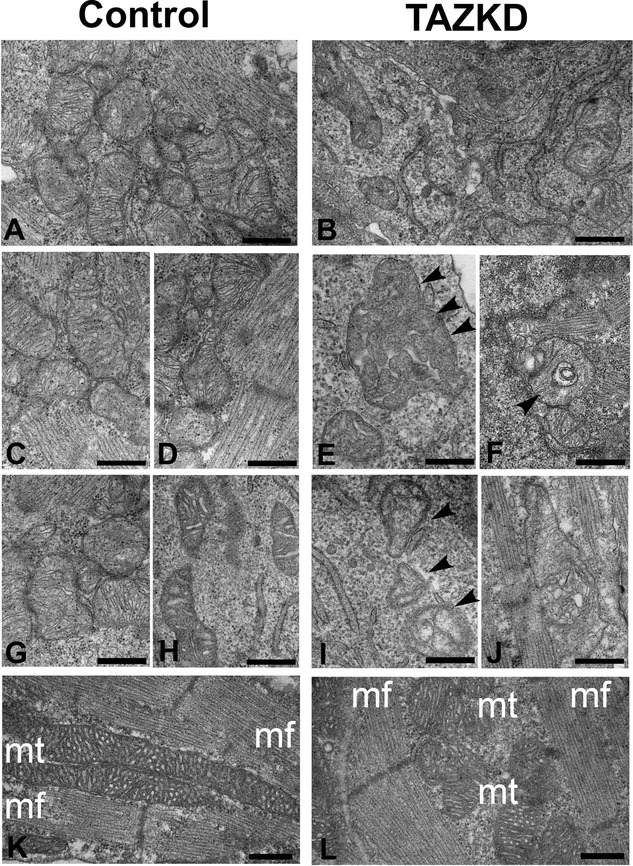
Electron micrographic analysis shows a spectrum of mitochondrial abnormalities similar to human Barth syndrome. We compared E13.5 embryonic wildtype (A, C, D, G, H) and TAZKD (B, E, F, I, J) cardiomyocytes, focusing on the mitochondria and regions around the mitochondria. Examples of abnormal mitochondrial ultrastructure include reduced mitochondrial density (A, B); abnormal cristae morphology (B, E, F, I, J), including disruption of cristae (B, E) and decreased cristae matrix density (I); and abnormally large mitochondria (E, J) (see arrowheads for selected abnormal mitochondria). Disruption of the normal alignment of mitochondria (mt) with myofibrils (mf) is shown in representative newborn control (wildtype) versus TAZKD cardiomyocytes (K, L). Scale bars: 500 nm. TAZKD indicates tafazzin knockdown.