
Figure 5.
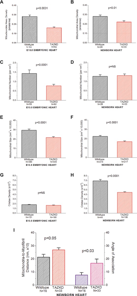
Morphometric analysis of mitochondria reveals distinct abnormalities in TAZKD mice. Mitochondrial structures were analyzed from electron micrographs taken of E13.5 embryonic hearts (A, C, E, G) and newborn hearts (B, D, F, H, I). TAZKD cardiomyocytes demonstrate statistically significant decreases in mitochondrial area density (embryonic, newborn), mitochondrial number per unit area (embryos), mitochondrial size (embryonic, newborn), and cristae density (newborns). Newborn myocardium exhibits disruptions in the spatial relations between mitochondria and myofibrils, as indicated by increased distances and a less-parallel geometry between mitochondria and myofibrils (I) (NB: Embryonic myocardium does not exhibit myofibrillar units well-developed enough for analysis). In A, B, C, F, and I, the N shown are the number of fields used for data analysis; in E, F, G, and H, N is the number of mitochondria analyzed. TAZKD indicates tafazzin knockdown.