
Figure 6.
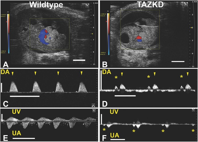
Cardiac dysfunction is evident in some TAZKD embryonic mice. Some TAZKD embryos exhibit severe diastolic dysfunction with a unique “atrial kick” in the dorsal aorta, at E12.5, 13.5, and 14.5, as revealed by spectral Doppler analysis of dorsal aortic blood flow; images are of an E13.5 wildtype embryo (A, C, E) and TAZKD embryo (B, D, F). Sagittal views of control wildtype (A) and TAZKD (B) embryos with superimposed color Doppler map show blood flow, but no obvious abnormalities such as hydrops (scale bars = 2 mm). Normal all-antegrade dorsal aortic (DA) blood flow is seen in the wildtype embryo (C), with peak aortic pulsations indicated by the arrowheads. Blood flow in the TAZKD dorsal aorta (D) shows an “atrial kick” (asterisks) occurring just before the peak aortic pulsations (arrowheads). In umbilical artery (UA) and umbilical vein (UV), the TAZKD embryo (F) shows propagation of the atrial kick even into the umbilical artery (asterisks), not evident in the wildtype embryo (E). Also note peak aortic velocities in TAZKD embryos (D, F) are lower than in WT littermates (C, E). Time (horizontal) scale bars for panels C to F = 0.5 s; velocity (vertical) scale bar = 100 mm/s. TAZKD indicates tafazzin knockdown.