
Figure 7.
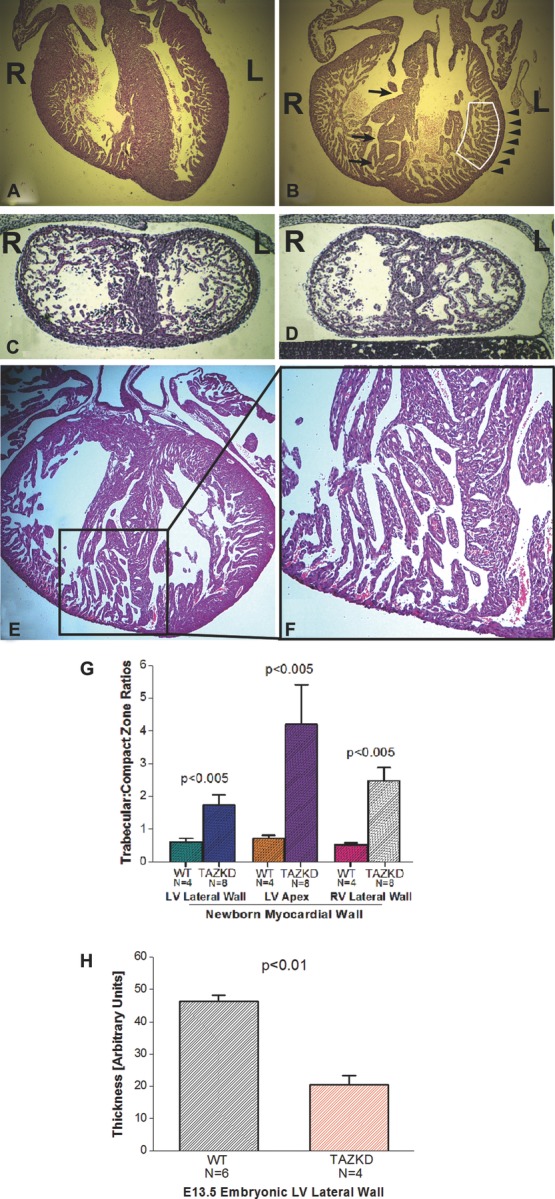
TAZKD mice exhibit thinner myocardium with prominent trabeculations suggestive of myocardial noncompaction. “4-chamber” views of newborn WT control (A) and TAZKD (B) hearts show deeper trabecular recesses and more abundant trabeculae (boxed area), thinner myocardium (arrowheads), and a less well-developed and more perforate interventricular septum in TAZKD hearts (arrows) (R = right, L = left). Coronal sections of E13.5 embryonic WT control heart (C) and TAZKD (D) hearts also demonstrate a thinner myocardium with more prominent trabeculae that occupy more of the left ventricular lumen in TAZKD hearts; again, the septum appears more porous. Tafazzin knockdown in these mice (A to D) was induced from start of gestation. Induction of TAZKD starting at E10.5 (E) recapitulates the cardiomyopathic phenotype, as represented by this newborn TAZKD heart (enlargement of apex in [F]). TAZKD newborn pups show a significantly higher degree of hypertrabeculation-noncompaction and myocardial thinning in all areas, but most notably the LV apex (G). In E13.5 TAZKD littermate embryos (H), significant myocardial thinning is also evident. Because this embryonic stage still exhibits a normally prominent trabecular myocardium, quantitation of trabecular-compact zone myocardium thicknesses was not performed. TAZKD indicates tafazzin knockdown; WT, wildtype.