
Figure 4.
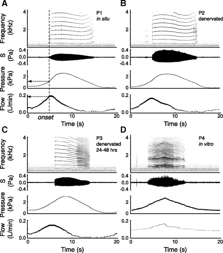
The spectrum complexity increases from in situ to an excised preparation. Typical run of each preparation, with sound spectrogram, sound amplitude, pressure and flow traces. A–D, Preparation P1, in situ phonation (A); preparation P2, in situ phonation after lesion of the bilateral tracheosyringeal branches of the hypoglossal nerve (B); preparation P3, in situ phonation 24–48 h after lesion (C); and preparation P4, excised in vitro preparation (D). A pressure ramp of 2 kPa gauge pressure was imposed in each case except for P4 where suction was applied on the trachea (compare with Fee et al., 1998). The dashed vertical line indicates the phonation onset for P1 with corresponding onset pressure and onset flow (arrows).