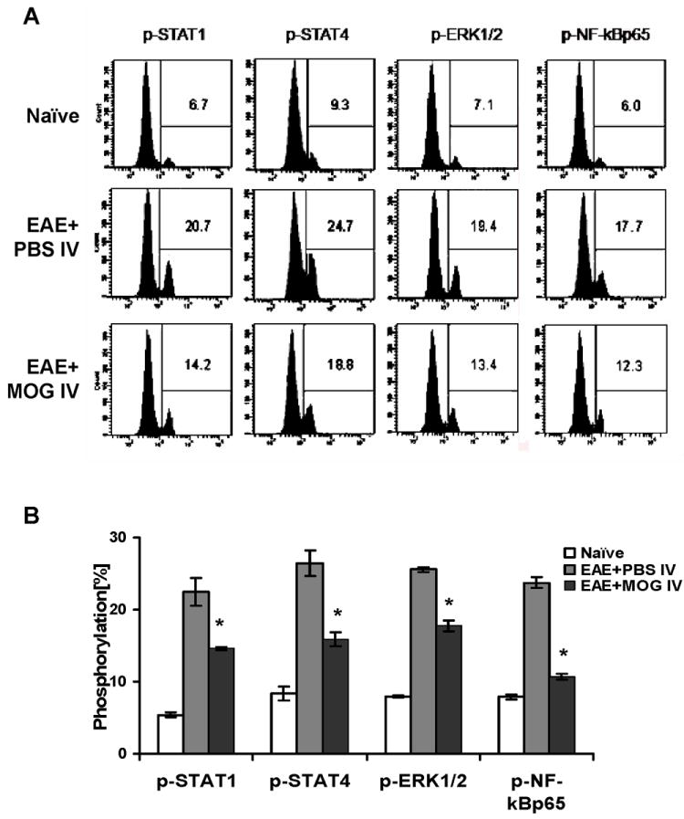
Fig. 3. MOG35 i.v. inhibited tyrosine phosphorylation of STAT4 and NF-κB in splenocytes.

2-3 × 10⁶ spleen cells from mice at day 21 p.i. were stimulated in vitro with 10 μg/ml MOG35-55 for 12 hrs. Cells were harvested and incubated with antibodies against p-STAT1, p-STAT4, p-ERK1/2, p-NF-κBp65 for flow cytometric analysis. One representative data set of 3 experiments is shown in (A). (B) Statistical analysis of phosphorylation in each group. Data were pooled from two independent experiments and presented as mean percentage of positive populations in splenocytes per group ± s.e.m. (n =10 each group). * p<0.05 as comparison between EAE + PBS i.v. and EAE + MOG i.v. mice.
