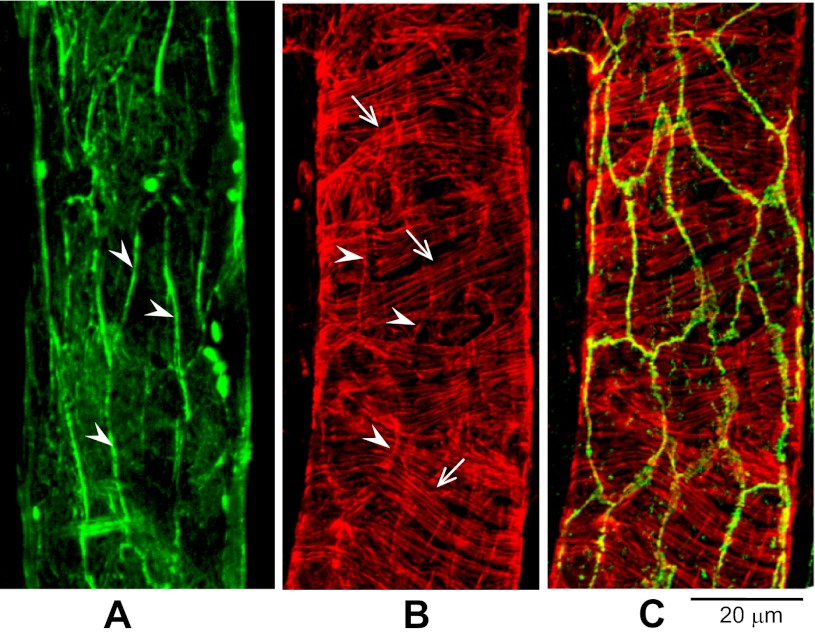
Fig. 3.
Peripheral F-actin colocalizes with VE-cadherin under control conditions in day 1 vessels. A: endothelial cell peripheral F-actin (green, arrowheads). B: F-actin staining (red) in both endothelial cells (arrowheads) and pericyte (arrows). C: the same vessel segment shown in B, but with double staining for F-actin (red) and VE-cadherin (green), demonstrating the overlap of VE-cadherin with endothelial F-actin peripheral bands.

