Figure 1.
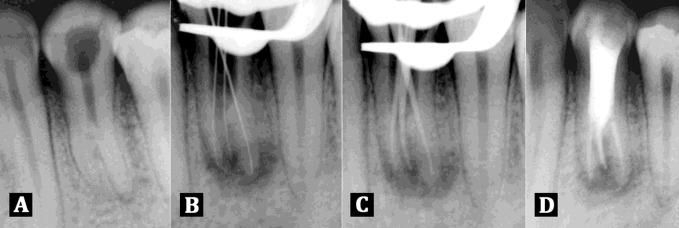
A) Diagnostic radiograph showing three roots inmandibular left first premolar; B) Working length radiograph of three rooted mandibular left first premolar was taken with size 10 K files; C) Master cone radiograph of three rooted mandibular left first premolar was taken with F1 protaper cones; D) Radiograph showing obturation of all the three canals of mandibular left first premolar

