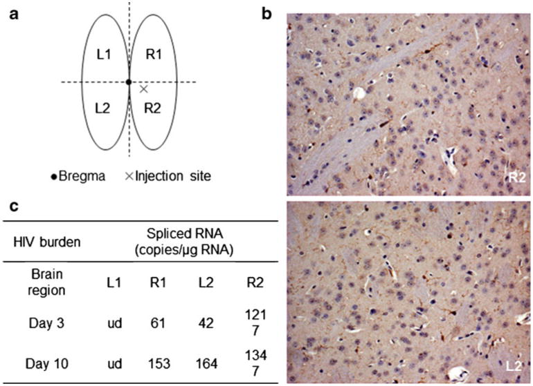
Fig. 2.

IC inoculation of Eco-HIV into mouse brain. a Schematic view of site of injection and dissected brain regions tested for HIV expression; b ICC for Iba-1 in the right basal ganglia region (R2) beneath and close to the injection site 3 days after IC inoculation of 10 μl of saline; note minimal microglial activation and absence of pathological damage. For comparison, ICC staining for Iba-1 in an analogous basal ganglia region from left (uninjected; L2) hemisphere from the same mouse. 10× magnification. c HIV expression proximal and distal to the injection site 3 days after IC inoculation with EcoHIV
