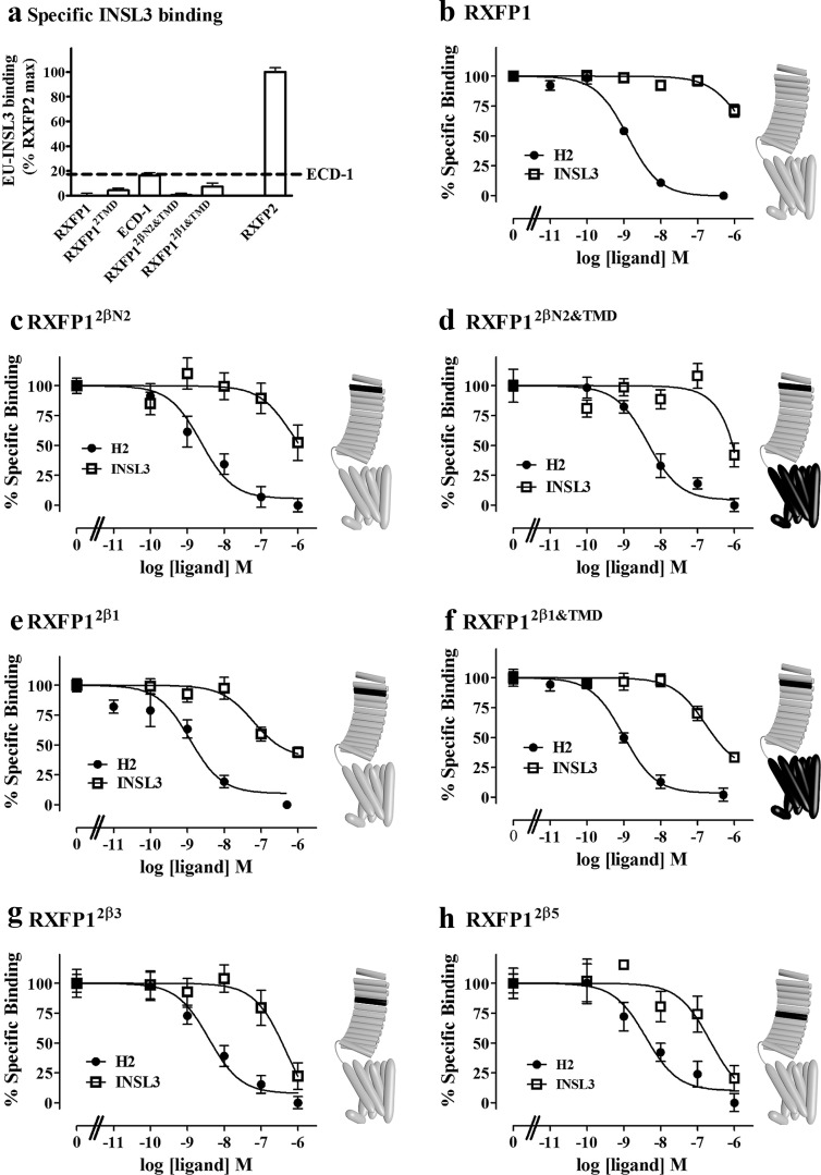
Fig. 3.
A, Eu-INSL3 binding to full-length chimeric receptors, normalized to that of RXFP2. Eu-H2 relaxin competition binding curves on (B) RXFP1, (C) RXFP12βN2, (D) RXFP12βN2&TMD, (E) RXFP12β1, (F) RXFP12β1&TMD, (G) RXFP12β3, and (H) RXFP12β5 expressed on the surface of cells. Eu-H2 relaxin was competed with either H2 relaxin (solid circles) or INSL3 (open squares). Schematic representations of the receptor chimeras are displayed to the right of relevant graphs. Gray regions indicate RXFP1 sequence with black regions representing the insertion of RXFP2 sequences.

