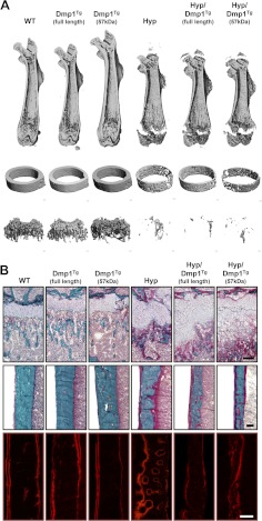
Fig. 4.
Bone phenotype of 5-wk-old WT, Dmp1Tg(full length), Dmp1Tg(57 kDa), Phex-deficient (Hyp), Hyp/Dmp1Tg(full length), and Hyp/Dmp1Tg(57 kDa) mice. A, 3D-μCT representation of (from top to bottom) the entire femur, the femoral midshaft, and the femoral distal metaphysis. B, Modified Goldner staining on femur histological section showing (top panels) the distal growth plate and metaphysis and (middle panels) the cortical bone at midshaft; the bottom panels show the fluorescent Alizarine Red S double labeling of the cortical bone. Scale bar represents 100 μm.

