
FIGURE 1.
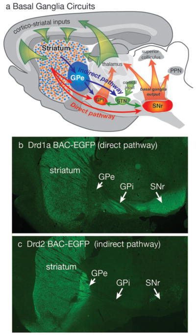
Diagram of basal ganglia circuits (a). The striatum receives excitatory corticostriatal and thalamic inputs. Outputs of the basal ganglia arise from the internal segment of the globus pallidus (GPi) and substantia nigra pars reticulata (SNr), which are directed to the thalamus, superior colliculus and pendunculopontine nucleus (PPN). The direct pathway originates from Drd1a expressing SPNs that project to the GPi and SNr output nuclei. The indirect pathway originates from Drd2 expressing SPNs that project only to the external segment of the globus pallidus (GPe), which together with the subthalamic nucleus (STN) contains transynaptic circuits connecting to the basal output nuclei. The direct and indirect pathways provide opponent regulation of the basal ganglia output interface. (b) Fluorescent imaging of a brain section from a mouse expressing EGFP under regulation of the Drd1a promoter shows Drd1a expressing SPNs in the striatum that project axons through the GPe, which terminate in the GPi and GPe. (c) Fluorescent of imaging of a Drd2-EGFP mouse shows that labeled SPNs provide axonal projections that terminate in the GPe, but do not extend to the GPi or SNr.