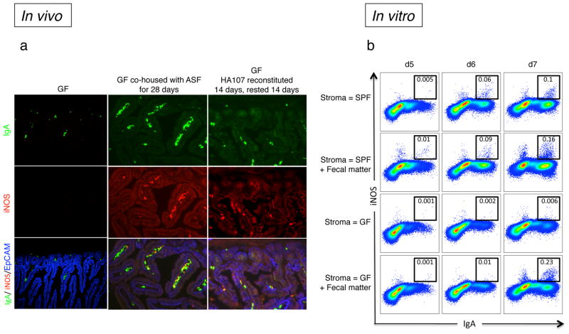
Figure 2. TNFα and iNOS-expression in IgA+ plasma cells requires microbial exposure.
a, Sections of small intestines of germ-free (GF) and GF mice that were re-colonized with either a defined Altered Schaedler Flora (ASF), or reversibly re-colonized with HA107 E. coli by continual gavage administration for 14 days followed by 14 days without gavage returning them to a GF status10. All sections were stained with specific fluorochrome-tagged antibodies for IgA (green), iNOS (red) and EpCAM (blue) and analyzed by fluorescence microscopy. Representative pictures are shown from 6 mice per group. b, B220+ bone marrow (BM) cells from CD45.1+ wild type mice were co-cultured for 7 days with CD45.2+ intestinal lamina propria cells (referred to as gut stroma) from either SPF or GF animals in the presence of IL-7, TGFβ, IL-21 and αCD40 antibody. In some cases faecal matter (intestinal wash) was added on the first day of the culture. The expression of IgA and iNOS was analyzed by flow cytometry, and the black boxes that indicate cells that are considered iNOS+ were based on the absence of staining derived from isotype controls (not shown). To ensure selective analysis of BM-derived precursors, cells were pre-gated on the CD45.2− population. Representative flow cytometry plots of cells are depicted. Data are representative of at least two independent experiments with 3–4 mice per group per experiment.

