
Fig. 2.
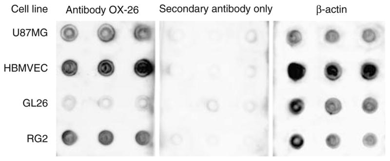
Dot blot analysis of species cross-reactivity of transferrin receptor antibody OX-26. Lysate of the rat glioma cell line RG2 (positive control) reacts well with OX-26. Mouse glioma GL26 cell lysate shows background reactivity (negative control). Lysates of human glioma U87MG and of human brain microvascular endothelial cells (HBMVEC) also react well with the antibody. Omission of primary antibody (secondary antibody; middle panel) only produced background signal. A blot for β-actin as positive loading control is shown on the right panel. Results are shown for 8 μl lysates in triplicates