Fig. 5.
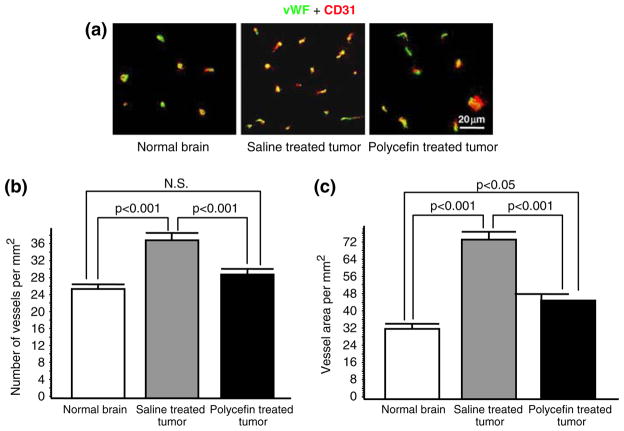
Decreased vascular density and area in tumors treated with Polycefin. (a) Double immunohistochemical staining of rat brain vessels using antibodies to two endothelial markers, von Willebrand factor (vWF, green) and CD31 (red). The two markers were used to optimize the screening accuracy. In most vessels both markers co-distribute (yellow color). The vessel number is higher in the xenotransplanted U87MG tumor than in normal brain, and this number is decreased after four intracranial Polycefin treatments. (b) Quantitative assessment of vascular density in treated and untreated tumors compared to normal brain. Vessels were revealed by either marker (Fig. 5a) and their number was quantitated at × 200 direct magnification. Images were analyzed using NIH ImageJ software. Statistical significance was determined by ANOVA. Microvascular density in xenotransplanted U87MG human glioma mock-treated with saline is significantly increased compared to normal brain (p < 0.001). After four intracranial treatments with Polycefin, tumor vessel density was significantly decreased (p < 0.001) and became similar to normal brain tissue (NS, not significant with p > 0.05). (c) Quantitative assessment of vascular area in treated and untreated tumors compared to normal brain. Vessels were revealed by either marker (Fig. 5a) and their relative area quantitated as for vessel density. Vessel area in xenotransplanted U87MG human glioma mock-treated with saline is significantly increased compared to normal brain (p < 0.001). After four intracranial treatments with Polycefin, tumor vascular area significantly decreased (p < 0.001) but remained somewhat higher than in normal brain (p < 0.05)

