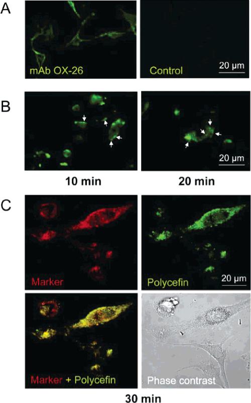
Figure 4.
Drug delivery into cultured human glioma U87MG cells. A. U87MG glioma cells display surface staining with OX-26 antibody by indirect immunofluorescence demonstrating cross-reactivity with human antigen (left). Omission of primary antibody abolished staining (negative control, right). B. Left, 10 min of U87MG treatment with Fluorescein-labeled Polycefin. The location of Polycefin is indicated by green fluorescence near the cell membrane (arrows), and early endosomes are beginning to form. Right, endosomal formation after 20 min treatment with Polycefin. Maturing endosomes are visible inside the cells (arrows). C. Co-distribution of endosomal marker FM 4-64 (marker) with Fluorescein-Polycefin (30 min) in cultured U87MG cells 30 min after treatment. FM 4-64 stains endosomes (red color), and Polycefin is found in the same place (green color). Co-localization is revealed as yellow color (lower left). Confocal microscopy.

