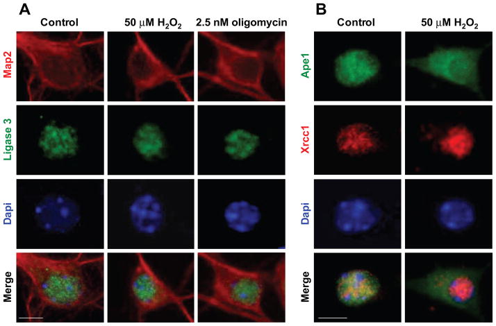
Figure 4. Nuclear localization of BER proteins, ligase 3 and Xrcc1 is maintained, while nuclear Ape1 is depleted following H2O2 and oligomycin exposures.
(A) Immunofluorescent images of DIV7 neurons reacted with anti Map2 antibody (red) and anti-ligase3 (green), show nuclear immunoreactivity of ligase 3, which coincides with DAPI (blue; scale bar=6 μm). Nuclear localization of ligase 3 (green, left panel) was not altered by either 50 μM H2O2 or 2.5 nM oligomycin exposures, and fully retained in the nuclear compartment (middle and right panels). (B) Xrcc1 is retained while Ape1 is depleted from nuclei and accumulates in cytoplasm following H2O2 exposure. Following exposure to H2O2, Xrcc1 immunoreactivity is retained in nucleus (red), while Ape1 immunoreactivity (green) shifts to cytoplasm (top, right); consequently, no overlap of Xrcc1 and Ape1 immunoreactivity is detected in merged images after treatment (bottom, right).

