
Figure 2 .
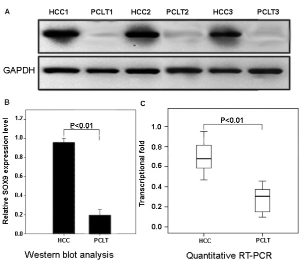
Increased expression levels of L1CAM protein and mRNA in hepatocellular carcinoma (HCC) and adjacent nonneoplastic liver tissues. (A) Representative Western blotting of L1CAM protein levels in HCC tissues and adjacent nonneoplastic liver tissues. (B) Semiquantitative Western blotting showed significantly increased L1CAM protein level in HCC tissues compared with adjacent nonneoplastic liver tissues. GAPDH was used as internal control. Means, standard deviation (SD), and P values were given (T test). (C) Significantly increased L1CAM mRNA level (P <0.01, Mann–Whitney test) in HCC tissues compared with adjacent nonneoplastic liver was detected by quantitative RT-PCR. GAPDH was used as internal control.