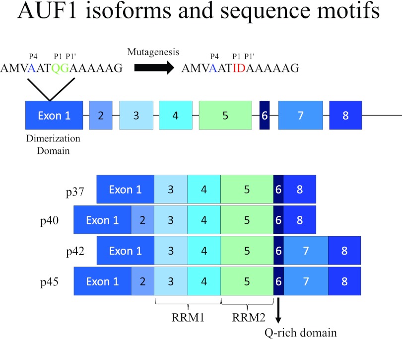
FIG 1.
AUF1 exon structure schematic and proposed 3CD cleavage site. The figure shows the exon structure of the human AUF1 gene and the different protein isoforms (p37, p40, p42, and p45) generated by alternative splicing. The RNA recognition motifs (RRM1 and RRM2), the Q-rich domain, and the dimerization domain are indicated. The top line of the figure shows an expansion of the amino acid sequence in exon 1 starting with amino acid residue 29 and extending through residue 42. This region contains a putative picornavirus 3CD cleavage recognition site with the P1 and P1′ positions Q-G (highlighted in green) preceded by an upstream A in the P4 position (highlighted in blue). For some of the experimental results displayed in Fig. 3, the Q-G pair was mutagenized to I-D (highlighted in red). This amino acid pair is predicted not to be cleaved by the poliovirus or human rhinovirus 3CD proteinase. Adapted with data from the work of Gratacos and Brewer (19).

