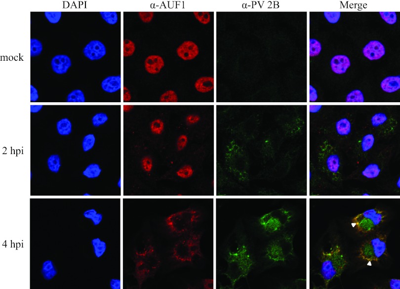
FIG 5.
Relocalization of AUF1 and partial colocalization with viral protein 2B during poliovirus infection. HeLa cells seeded on coverslips were either mock infected or infected with poliovirus and fixed at 2 or 4 hpi. Cells were immunostained with anti-AUF1 antibody (shown in red) or anti-poliovirus 2B antibody (shown in green) and stained with DAPI to visualize the nucleus (shown in blue). Colocalization of AUF1 and 2B, indicated in the merged image by white arrowheads, was examined by confocal microscopy.

