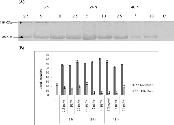
Figure 4.

(A) Changes in expression of apoptotis-related protein in response to treatment with digallic acid. TK6 cells were treated with 2.5, 5 and 10 μg/ml of digallic acid for 6, 24 and 48 h. Protein extracts were subjected to western blotting to determin immunoreactivity levels of PARP, as described in methods section. PARP 116 KDa and 85 KDa bands are shown. C: Cells treated with 0.5% DMSO. (B) Quantification by scanning densitometry of PARP bands intensity*.
