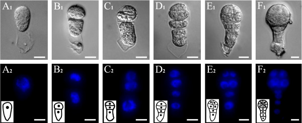
Figure 1 .
Cell division pattern of zygote and proembryo in tobacco. (A1-H1) Bright-field images. (A2-H2) Fluorescence images of DAPI-stained nuclei corresponding to bright-field images. (A1 and A2) A isolated live zygote at 84 h after pollination. (B1 and B2) A two-celled proembryo with a small apical cell and a large basal cell from a zygote asymmetrical division. (C1 and C2) A three-celled proembryo with a large basal cell and two-celled embryo-proper after apical cell division. (D1 and D2) A four-celled proembryo with a two-celled embryo-proper and a two-celled suspensor. (E1 and E2) A proembryo with an eight-celled embryo-proper and a two-celled suspensor. (F1 and F2) A multicellular proembryo with a four-celled suspensor. The inserted pictures in A2-F2 are schematic diagrams of zygotes and proembryos. Bars = 10 μm.

