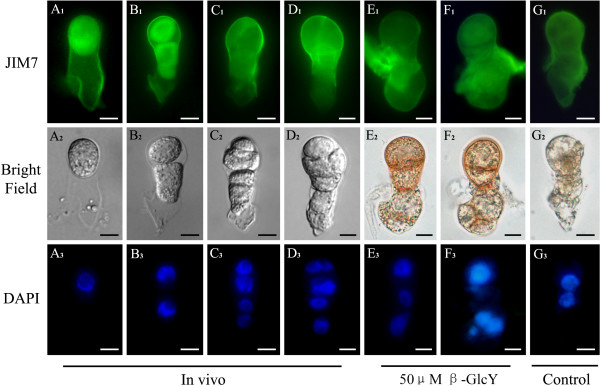
Figure 5 .
Distribution of high-esterified pectins by immunolabeling with JIM7 in zygotes and proembryos. (A1-G1) The fluorescence images of immunolabeling with high-esterified pectins antibody JIM7 under a fluorescence microscope. The immunofluorescence gradually localizes in the apical end of cell wall during zygotes divide into proembryos in vivo (A1-D1). In in vitro samples, high level immunofluorescence distributes in the whole cell walls of proembryos cultured with 50 μM β-GlcY (E1-F1) and the samples cultured without β-GlcY (G1). (A2-G2) Bright-field images of zygotes and proembryos corresponding to A1-G1. (A3-G3) Fluorescence images of DAPI-stained nuclei corresponding to bright-field images. Bars = 10 μm.

